The Shroud of Turin—Physical Description
Wojciech Kucewicz ![]()
AGH University of Science and Technology, Kraków, Poland
Jakub S. Prauzner-Bechcicki ![]()
Jagiellonian University in Krakow, Poland

In order to present such an intriguing object as the Shroud of Turin, it is necessary to organise the data collected over more than 100 years of research. The description of the Shroud can be divided into three categories:
- the description of the material from which the Shroud was made,
- the description of the traces present on the Shroud, and
- the description of the image.
This is a very synthetic characterisation. For references to detailed research results, see: →Physical Analyses of the Shroud, and →Chemical Analyses of the Shroud. Illustrative material is also included showing: (A) a photograph of the Shroud, (B) the same image with contrast correction to emphasise detail, and (C) a diagram showing the main elements visible on the Shroud. The photograph of the Shroud (A) is cropped, i.e. the Shroud is not visible in its entirety: for example, the part with the frontal image is slightly shorter than in reality. The diagram (C) uses different coloured lines to mark different types of objects visible on the Shroud: the grey line marks the contours of the image, the red line marks some of the traces of blood and wounds, the blue line marks selected spots created after extinguishing with water, the black line marks the contours of the burns, and the orange line marks the holes burnt in 1532.
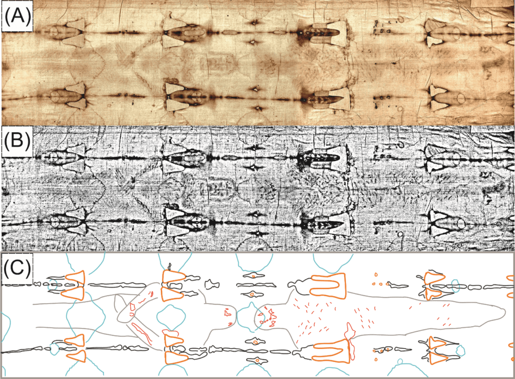
(B) The same photo in a monochromatic version, with additional contrast adjustment
(C) Diagram showing the image of the figure and the most significant marks visible on the Shroud
Material
The Shroud was made of linen fabric measuring 4.3 × 1.1 m (Schwalbe, Rogers 1982). The flax yarn comes from the flax stem, hence the main component is cellulose. Aging of the cellulose causes yellowing of the material (Schwalbe, Rogers 1982—see item 43 in the list of references from this work). The fibres have a Z-shaped twist (Schwalbe, Rogers 1982; Raes 1991). The threads of the fabric have a diameter of 0.15 mm and are formed from fibres with diameters of 10–15 µm (Schwalbe, Rogers 1982).
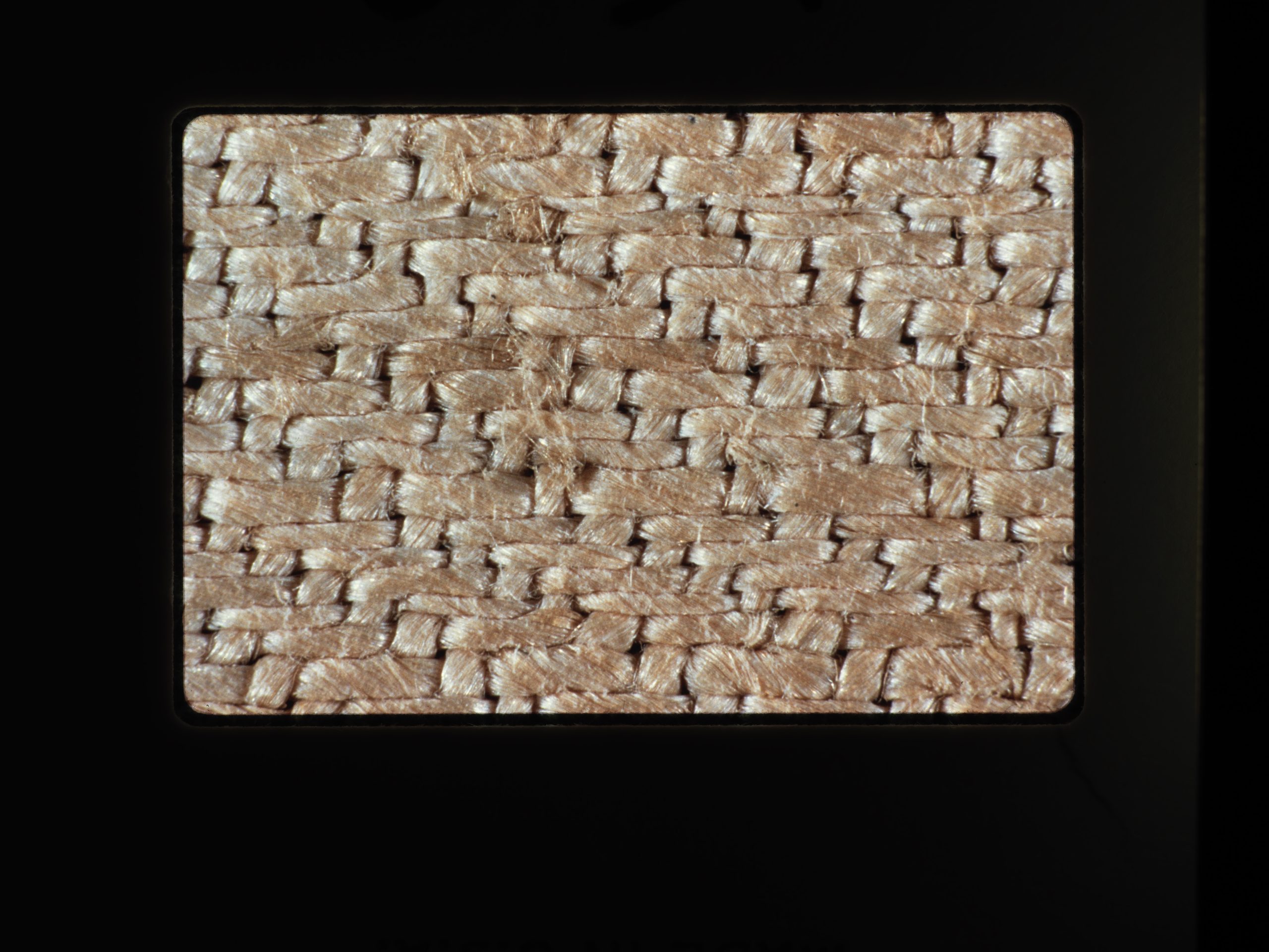
The fabric was created by interlacing warp and weft threads on the loom, creating a 3:1 twill weave (known as herringbone weave), i.e. the weft thread passes alternately under three and over one warp thread (Schwalbe, Rogers 1982; Wilson 1985). At the time of Christ, linen cloth was woven using a plain weave, in which the weft thread passes alternately above and below the warp thread. The herringbone twill weave makes the fabric more expensive (Wilson 1985). However, it is not a feature that would make it easier to determine the age or place of origin of the fabric (Schwalbe, Rogers 1982; Raes 1991).
In addition to the weave, there are two other important facts to be considered in relation to the way the fabric of the Shroud was woven. First, the examination of the samples taken did not reveal the presence of fibres of animal origin (i.e. wool or silk), but did reveal the presence of cotton fibre (Gossypium herbaceum) (Schwalbe, Rogers 1982; Raes 1991). This leads to the conclusion that the fabric was created on a loom on which only fibres of plant origin were woven, which clearly refers to Jewish law, which forbade the use of the same loom for weaving animal and plant threads (Wilson 1985; Górny, Rosikoń 2012).
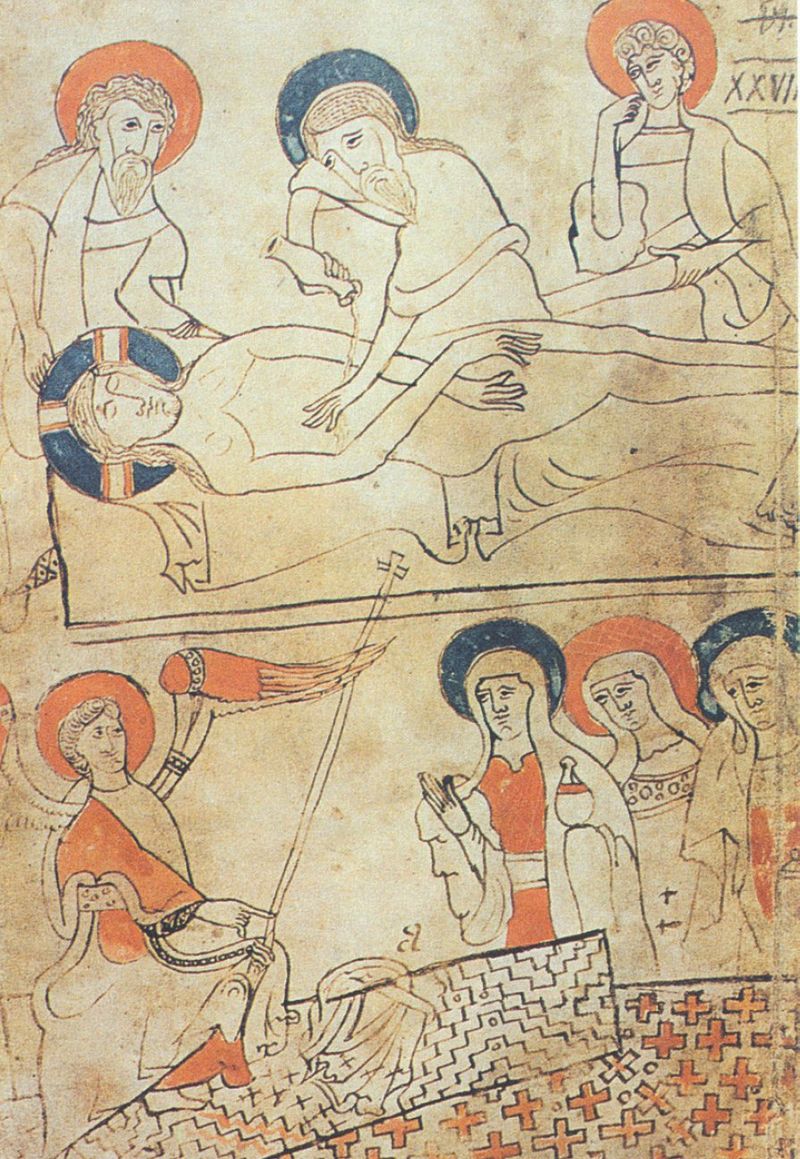
Secondly, the distinctive weave seen on the Shroud was reproduced in miniatures depicting the linen in which Jesus was wrapped, included in the oldest document known today written in Hungarian, the so-called Pray Codex. This document, held in the Széchényi National Library in Budapest, is dated between 1192 and 1195 (almost 70 years earlier than the age of the Shroud as determined by a study using radiocarbon dating, →Determination of the Age of the Shroud), and was discovered by György Pray, a priest, in 1770 (Wilson 1985; Górny, Rosikoń 2012; Marion, Lucotte 2008). Along the longer edge of the linen on which the image is visible (Schwalbe, Rogers 1982), an identical-looking narrow strip of a width varying between 7.8 and 8.4 cm is sewn on. The seam connecting the two parts is 4–5 mm wide. It has not been established with certainty whether the main and the sewn-on parts are of different origin (Schwalbe, Rogers 1982; Raes 1991).
In 1532, a fire damaged the silver reliquary in which the Shroud was stored. Drops of molten metal burned holes in the fabric, over which the Poor Clares of Chambéry sewed patches, and the Shroud was lined with Dutch linen (Pellicori, Evans, 1981). In 1978, during the →STURP work, the linen protecting the reverse of the Shroud, which appeared brighter than the obverse, was partially removed (Schwalbe, Rogers 1982). The complete removal of the Dutch linen was done in 2002 during conservation work, and a brand-new linen was sewn to the reverse (Resch 2006).
Traces
There are a number of clearly visible traces on the Shroud that are not directly associated with the image (see Fig. 1). These include traces of burning, including those evidenced by the fire of la Sainte-Chapelle de Chambéry in France in 1532, and those made on other occasions, in addition to traces made by extinguishing water (Pellicori, Evans, 1981; Wilson 1985; Górny, Rosikoń 2012; Marion, Lucotte 2008; Resch 2006). The latter are visible as regularly recurring stains, streaks [see the contours of the stains marked with the blue line in figure (C)]. Their regularity is related to the way the fabric was folded during the extinguishing. The burn marks associated with the 1532 fire are also characterised by a certain regularity [orange and black lines in figure (C)]. On the one hand there are two parallel brown lines extending along the length of the cloth, while on the other hand there are lines of four triangular holes occurring four times at similar intervals and two additional sets of four damaged spots at the two shorter ends of the cloth. The regularity of the occurrence of these damaged spots also relates to the way the Shroud was folded to be placed in a silver reliquary during the fire. Other, less regular, traces of urning are also visible (Schwalbe, Rogers 1982; Wilson 1985). According to →STURP researchers, these traces can most likely be linked to the dripping of flammable material from the torch or flambeau with which the Shroud was illuminated (Schwalbe, Rogers 1982). Reproductions of these traces can also be seen in the miniatures contained in the Pray Codex, which, given the age of this document, would suggest that they were made before the fire of 1532.
Image
What is of greatest interest is the image of the figure visible on the surface of the Shroud of Turin: the frontal and rear images [the approximate contours of the figure are marked with the grey line in the figure (C)]. Due to the slight contrast between foreground and background, the image is faint, best seen from a certain distance (4–5 m) (→Photographs of the Shroud). Nevertheless, it is possible to discern with certainty on the Shroud the figure of a man about 1.78 m tall and weighing 75–80 kg (Pellicori, Evans, 1981). →STURP researchers led by John Jackson have shown experimentally that the degree of shading in the image is a function of the distance between the cloth and the body that was wrapped in it (Jackson, Jumper, Ercoline 1982; Jackson, Jumper, Ercoline 1984; Ercoline, Downs, Jackson 1982). This relationship makes it possible to reproduce a three-dimensional image of the figure wrapped in the Shroud (Jackson, Jumper, Ercoline 1982; Jackson, Jumper, Ercoline 1984; Ercoline, Downs, Jackson 1982; Devan, Miller 1982; Bevilacqua et al. 2018), which is an astonishing property with regard to images created on the surface of any material. What is puzzling is that this three-dimensionality of the image is encoded only in the frontal view, while the rear image subjected to a similar procedure presents itself as flat.
The image, which is visible to the naked eye, was created by the yellow discolouration of the cellulose fibres (→Chemical Analyses of the Shroud) (Schwalbe, Rogers 1982; Jumper et al. 1984). In-depth analyses of the area of the canvas on which the image is located have shown that the fibres forming the image are discoloured only on the surface (→Hypotheses of the Origin of the Image on the Shroud) (Jumper et al. 1984; Fanti et al. 2005; Fanti 2014; Fanti et al. 2010a; Fanti et al. 2010b). The image has a chiaroscuro characteristic, i.e. the darkening depends on the number of discoloured fibres per unit area. The shade of the discoloured fibres is the same in the bright and dark areas of the image. The tests did not show the fibres to be bonded or that there is capillary flow of fluids in the image area. There are also no pigments or brush marks. In the areas damaged by the 1532 fire, no evidence was found of burnt paint binders from which the image would have been made. One would expect more rapid burning of protein binders, based on vegetable gum or starch, than of the cellulose contained in linen (Schwalbe, Rogers 1982). All this together strongly invalidates the hypotheses of the artistic origin of the image (→Hypotheses of the Origin of the Image on the Shroud).
The image of the figure is clearer in the photographs, and reveals a puzzling property: the negative of the photograph shows a positive image. In other words, the Shroud shows the kind of image we would observe on photographic film (→Photographs of the Shroud).
The figure visible on the Shroud has traces of blood and many wounds, the appearance and distribution of which on the body strongly indicate that the man depicted was subjected to cruel and deadly torture: he was scourged, had his head crowned with a cap (crown) of thorns, was beaten on the face, was forced to carry a heavy object on his back, was nailed to a cross and pierced in the side with a sharp, long instrument (→Exegetical and Medical Study of the Shroud). Traces of this kind are marked with a red line in the figure (C).
The →STURP team, approaching the research with due care, described the areas that looked like bloodstains quite accurately, and concluded that they were indeed bloodstains (Schwalbe, Rogers 1982). The results at the time, and subsequent observation, lead to the conclusion that the areas of the bloodstains and the image itself appear quite different in colour, texture and composition. In contrast to the image, which is faint, the bloodstains are very distinct. In reflected light they are visible as brownish-red, and in transmitted light—as scarlet/crimson. Under a magnification of 50 times, those stains look as if a viscous liquid was used to create them, which flowed around the thread and soaked into the fabric, leaving visible spots on the other side of the fabric as well. The characteristic features of the meniscus that accompanies the flow of viscous fluid can be seen, and the threads are clumped together (Schwalbe, Rogers 1982).
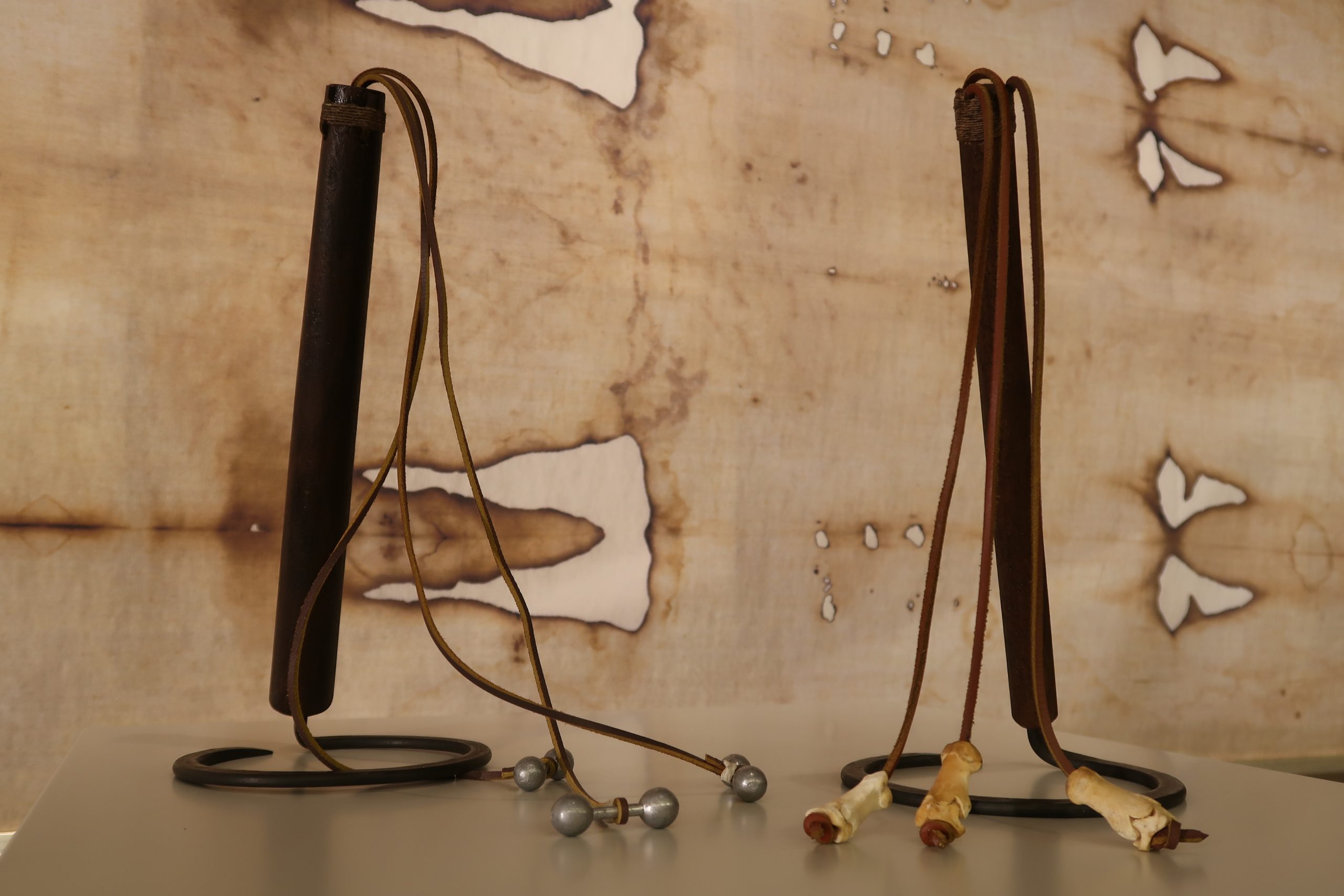
The most numerous are traces of small, three-centimetre-long wounds, the shape and distribution of which correspond to the flagellation performed with the Roman flagrum. This tool consisted of a wooden handle with attached thongs ending in small lead balls or pieces of bone. There are more than 120 such marks. The arrangement of the wounds on the body, front and back, suggests that there were two executioners, and the number of wounds would indicate that the man was sentenced to the Roman punishment of whipping, as Jewish law limited the number of lashes (Wilson 1985; Górny, Rosikoń 2012; Marion, Lucotte 2008).
Next, numerous traces of blood can be seen from the stab wounds to the head, the number and location of which indicate that something like a spiked cap was placed on the condemned man’s head (Pellicori, Evans, 1981; Wilson 1985; Górny, Rosikoń 2012; Marion, Lucotte 2008). The distribution of the traces of blood visible on the head of the figure depicted on the Shroud made it possible to make a reconstruction of the crown of thorns: in shape it resembles a spiked helmet, the wearing of which must have been extremely painful. There are also traces of torture on the face which are not so clearly visible to the naked eye (Pellicori, Evans, 1981; Wilson 1985; Górny, Rosikoń 2012; Marion, Lucotte 2008). The work of →STURP members and high-resolution photographs reveal a torn right eyelid, swollen nose, swollen cheeks and eyebrow arches. The image of the back, at the level of the right shoulder and both shoulder blades, shows abrasion marks resulting from contact with a heavy and angular object (Pellicori, Evans, 1981; Wilson 1985; Górny, Rosikoń 2012; Marion, Lucotte 2008). This would correspond to the description of carrying a cross. Such a cross consisted of a vertical beam called stipes and a horizontal beam called patibulum. In all likelihood, it was the latter that the condemned man carried on his own shoulders.
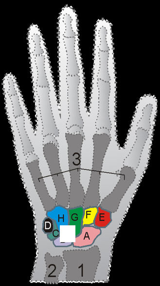
Other marks connected with the crucifixion of the figure depicted on the Shroud are those left by the nails piercing the hands and feet and by the blood flowing from these wounds. The piercing of the hands occurred in the area of the wrists in a place called Destot’s space, as evidenced by the clear presence of blood oozing from the area of the left wrist. This location of the nails ensured that the condemned man would not fall off the cross and thus it prolonged his suffering. Puncturing the hand in this particular location can damage the median nerve, resulting in thumb contracture (Bevilacqua et al. 2014). This is also how the image appears on the Shroud, where only four fingers are visible in the hand. Among the Pray Codex miniatures mentioned, there is one depicting the preparation of Jesus’ body for burial, and only four fingers are visible in the hand: the same number of visible fingers as in the figure on the Shroud (Górny, Rosikoń 2012; Marion, Lucotte 2008).
The wounds on the wrists explain the traces of blood on the forearms, while their positioning shows the direction of the blood flow and therefore also shows the positioning of the body at the time of the crucifixion (Bevilacqua et al. 2014). It can be concluded that, in trying to breathe in, the crucified man had to rise, supporting himself on his feet nailed to the cross, and then he was forced to fall, hanging on his hands. Traces of blood on the feet are only visible on the right foot in the front image and on both feet in the rear image. This distribution of stains suggests that the condemned man’s feet were attached to the cross by a single nail (Bevilacqua et al. 2014; Caja, Boi 2018).
Now the wound from the right-side puncture. The frontal image clearly shows that the puncture occurred between the fifth and sixth ribs. The size of the wound indicates that the impact was made with a spear and that it led to a puncture of the pleura, the pericardium and the right atrium of the heart, leading to profuse bleeding and leakage of plasma. The bloodstain created in the front image is very clear and concentrated around the wound, adjacent to the area damaged by the 1532 fire. In the rear image, the bloodstain extends across the entire width of the back and is situated between the damage from the fires. It is also accompanied by a stain from plasma leakage.
Particularly noteworthy is the fact that all the wounds, mutilations and accompanying traces of blood visible in the image of the figure from the Shroud correspond amazingly consistent with the descriptions of the Passion in the canonical Gospels (→Exegetical and Medical Aspects of the Death of Jesus).
References
Bevilacqua M. et al., How Was the Turin Shroud Man Crucified?, “Injury” 2014, Vol. 45, Supplement 6, pp. S142–S148, https://doi.org/10.1016/j.injury.2014.10.039.
Bevilacqua M. et al., Rigor Mortis and News Obtained by the Body’s Scientific Reconstruction of the Turin Shroud Man, “Forensic Science Today” 2018, 4(1), pp. 001–008, https://doi.org/10.17352/pjfst.000010.
Caja V.L., Boi M., The Evidence of Crucifixion on the Shroud of Turin Through the Anatomical Traits of the Lower Limbs and Feet, “Archaeometry” 2018, Vol. 60, No. 6, pp. 1377–1390, https://doi.org/10.1111/arcm.12383.
Devan D., Miller V., Quantitative Photography of the Shroud of Turin, [in:] IEEE Proceedings of the International Conference on Cybernetics and Society, 1982, [on-line:] https://www.shroud.com/pdfs/Quantitative%20Photography%20Devan%20Miller%201982%20OCRsm.pdf – 17 VII 2022.
Ercoline W.R., Downs Jr. R.C., Jackson J.P., Examination of the Turin Shroud for Image Distortions, [in:] IEEE Proceedings of the International Conference on Cybernetics and Society, 1982, [on-line:] https://www.shroud.com/pdfs/Examination%20for%20Image%20Distortions%20Ercoline%201982%20OCR.pdf – 17 VII 2022.
Fanti G. et al., Evidences for Testing Hypotheses about the Body Image Formation of the Turin Shroud, [in:] The Third Dallas International Conference on the Shroud of Turin, Dallas, 2005, [on-line:] http://www.shroud.com/pdfs/doclist.pdf – 29 X 2021.
Fanti G. et al., Microscopic and Macroscopic Characteristics of the Shroud of Turin Image Superficiality, “Journal of Imaging Science and Technology” 2010a, Vol. 54, No. 4, https://doi.org/10.2352/J.ImagingSci.Technol.2010.54.4.040201.
Fanti G. et al., List of Evidences of the Turin Shroud, [in:] Proceedings of the International Workshop on the Scientific Approach to the Acheiropoietos Images, ENEA, Frascati, Italy, Frascati 2010b.
Fanti G., Hypotheses Regarding the Formation of the Body Image on the Turin Shroud: A Critical Compendium, “Journal of Imaging Science and Technology” 2014, Vol. 55, No. 6, pp. 1–14, https://doi.org/10.2352/J.ImagingSci.Technol.2011.55.6.060507.
Górny G., Rosikoń J., Świadkowie tajemnicy. Śledztwo w sprawie relikwii Chrystusowych, Warszawa 2012.
Jackson J.P., Jumper E.J., Ercoline W.R., Three-Dimensional Characteristic of the Shroud Image, [in:] IEEE Proceedings of the International Conference on Cybernetics and Society, 1982, [on-line:] https://www.shroud.com/pdfs/3D%20Characteristic%20Jackson%20Jumper%201982%20OCR.pdf – 20 X 2021.
Jackson J.P., Jumper E.J., Ercoline W.R., Correlation of Image Intensity on the Turin Shroud with the 3-D Structure of a Human Body Shape, “Applied Optics” 1984, Vol. 23, No. 14, 2244, https://doi.org/10.1364/ao.23.002244.
Jumper E.G. et al., A Comprehensive Examination of the Various Stains and Images on the Shroud of Turin, “Archaeological Chemistry – III” 1984, 1, pp. 447–476, https://doi.org/10.1021/ba-1984-0205.ch022.
Marion A., Lucotte G., Tunika z Argenteuil i Całun Turyński. Podsumowanie badań, przeł. A. Łatka, Kraków 2008.
Pellicori S.F., Evans M.S., The Shroud of Turin Through the Microscope, “Archaeology” 1981, No. 34(1), pp. 34–43, [on-line:] https://www.shroud.com/pdfs/Shroud%20Thru%20Microscope%20Pellicori%20Evans%201981%20OCRsm.pdf – 17 VII 2022.
Raes G., The Textile Study of 1973-1974, “Shroud Spectrum International” 1991, No. 38/39, p. 3, [on-line:] https://www.shroud.com/pdfs/ssi3839part3.pdf – 24 III 2022.
Resch A., Oblicze Chrystusa. Od Całunu Turyńskiego do Chusty z Manoppello, przeł. A. Kuć, Radom 2006.
Schwalbe L.A., Rogers R.N., Physics and Chemistry of the Shroud of Turin, “Analytica Chimica Acta” 1982, Vol. 135, No. 1, pp. 3–49, https://doi.org/10.1016/S0003-2670(01)85263-6.
Wilson I., Całun Turyński, przeł. z ang. J. Piątkowska, Kraków 1985.
Sources of Images
1. Collection of W. Kucewicz and J.S. Prauzner-Bechcicki
2. Sindonology.org, Photomicrographs, http://www.sindonology.org/photomicrographs.shtml
3. Wikimedia Commons, https://commons.wikimedia.org/wiki/File:Hungarianpraymanuscript1192-1195.jpg (public domain)
4. Fragment of the exhibition “Who is the Man from the Shroud?” at the John Paul II Centre in Krakow. Collection and ownership of the Polish Syndonological Centre in Krakow
5. Wikimedia Commons, https://commons.wikimedia.org/wiki/File:Carpus.png (Zoph, CC BY-SA 3.0)