Blood on the Shroud
Jan S. Jaworski ![]()
University of Warsaw, Poland

The traces of blood on the Shroud of Turin, clearly visible to the naked eye and in photographs, have been studied from the very beginning. At first, scientists, having access only to photographs, compared the traces of blood with the descriptions of forensic specialists (→Exegetical and Medical Aspects of Jesus’ Death and Their Coherence with the Shroud). The expert report made by Pierre Barbet and presented in 1950 at the First International Congress in Turin turned out to be a milestone which confirmed that all the details were consistent with the knowledge he had gained from medical practice. Later, when the public exhibition of the Shroud took place, some scientists were able to examine the traces up close. Finally, from 1973 onwards, small samples taken from the linen were studied, and in 1978, the Shroud of Turin Research Project (→STURP) made direct examinations of the relic, including traces of blood. Since then, microscopic fragments remaining on the adhesive tapes pressed onto the linen in 1978 have been analysed in various laboratories, as well as material collected ten years later when samples were taken for radiocarbon testing of the age of the linen. Some of these samples were not returned to the Shroud’s owners, but—passed on to subsequent researchers—were studied until recent years. Neither the samples nor these examinations have the authorisation of the relevant scientific commission appointed by the Shroud’s owner, whose representative is the Archbishop of Turin. Caution should be exercised in generalising conclusions drawn from the examination of microscopic fragments of fibres and concerning traces of blood elsewhere on the cloth.
The scientific study of the traces of blood on the Shroud was aimed at finding answers to several fundamental questions. Are these traces of authentic blood? To which blood group do they belong? Is it human blood? How did the different blood traces originate from different types of wounds? Is it possible to distinguish between blood flowing from wounds during life and after death? Why has the blood on the Shroud retained its bright red colour over the centuries? What is the chemical composition of the blood residue on the Shroud and can it confirm the torture indicated by the wounds visible on the image? Can the preserved traces of blood indicate the age of the Shroud? Is the blood on the cloth of the Shroud consistent with the burial customs of the Jews at the time of Jesus?
The authenticity of the blood on the Shroud was established in the early 1980s by American research carried out as part of the STURP project and—independently—in Turin, which was subsequently confirmed by still other scientists. The electron spectra of blood traces recorded directly on the Shroud were shown to be similar to those of methaemoglobin (Gilbert and Gilbert 1980; Pellicori 1980; Heller and Adler 1980; Di Lazzaro 2015 team), the higher surface concentration of iron in areas of the blood traces than on the whole cloth and the decrease of this concentration as one moves away from the blood trace (Morris, Schwalbe and London 1980), the presence of a porphyrin ring, a basic chemical structure in the haemoglobin molecule (Heller and Adler 1980; Baima Bollone, 1980s), and the presence of albumin, a plasma protein (Heller and Adler 1981; Baima Bollone, 1980s), and bile pigments formed during the breakdown of haemoglobin: bilirubin (Heller and Adler 1981) and a mixture of biliverdin and bilirubin (Laude and Fanti 2017), as well as the fluorescence of the borders around the blood stains, indicating the presence of blood plasma (Miller and Pellicori 1981), the formation of the characteristic brown crystals of Teichmann-Stawiarski haemin (haemin hydrochloride) when acetic acid and sodium chloride crystals were added to the Shroud sample (Baima Bollone 1980), the solubility of the red coating of flax fibers and detached microscopic fragments in a mixture of certain enzymes and also in hydrazine with the formation of a pink colour characteristic of heme (Heller and Adler 1981), the formation of a bright red colour characteristic of cyanomethemoglobin after the addition of cyanides (Heller and Adler 1981), the detection by X-ray spectroscopy of elements characteristic of blood: iron from haem, chlorine and potassium from blood cell electrolytes, sulphur from blood plasma proteins, calcium, magnesium, aluminium and silicon (Baima Bollone 1981; Lucotte 2015; Fanti and Zagotto 2017) and the presence of antibodies belonging to immunoglobulin class G (Baima Bollone and Gaglio 1984).
In December 1982, Pierluigi Baima Bollone and his colleagues concluded by immunofluorescence that a sample of blood they had taken in 1978 flowing from the pierced side on the Shroud belonged to Group AB. This result was to be independently confirmed by M. Canale, while the American researchers, who had too little material, failed to confirm it. In 1985, P. Baima Bollone and his team also detected M, N and S antigens on a venous blood sample taken from the sole of the right foot on the Shroud, confirming that it was human blood, since S antigens are only characteristic of humans (and are not found in other primates like A and B antigens).
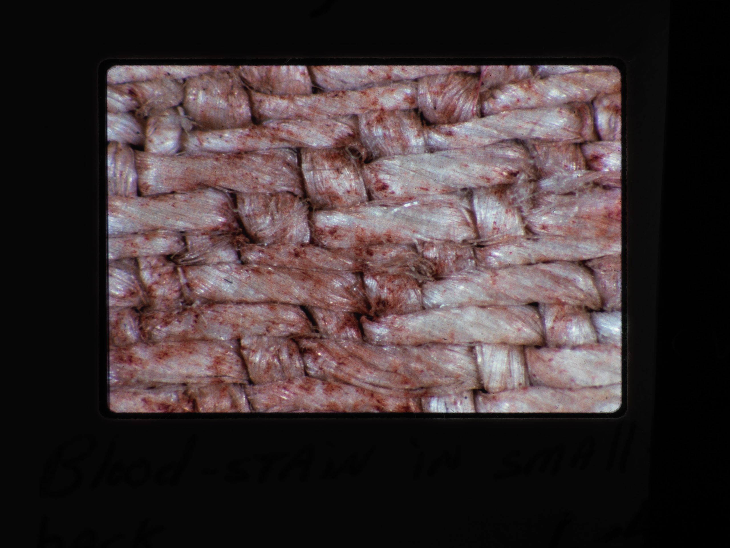
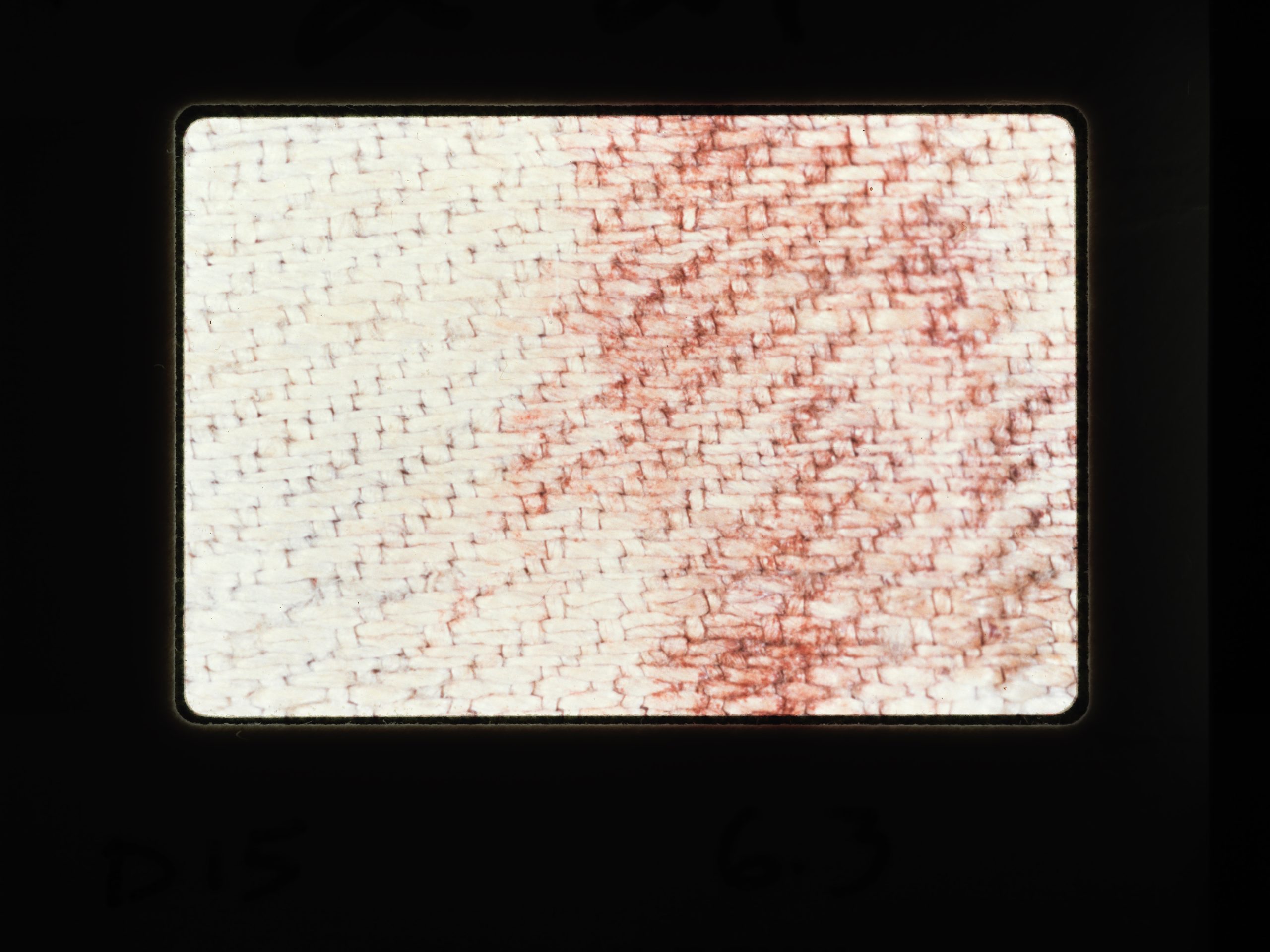
Forensic specialists, by analysing the wounds visible in the image and the blood flowing from them, were able to distinguish between venous and arterial blood and blood flowing during the ordeal and that flowing after death when the body was carried to the grave. Blood from different wounds soaked differently into the linen or formed droplets and clots and dissolved due to fibrinolysis, which resulted in differences in their physico-chemical properties. Chromatic analysis of the blood traces, an instrumental reading of the red colour at a specific wavelength, made it possible to divide the blood traces on the Shroud into six different areas, where the traces were formed in a slightly different way (Bedon et al. 2015).
The dispute over the presence of the painting pigments of brown ochre (iron oxide) and red cinnabar (mercury sulphide) in the areas of blood traces on the Shroud, which had been ongoing for several decades, reverberated widely in the syndonological literature. Opponents of the Shroud’s authenticity have argued that these were used to paint the blood marks and the image (McCrone 1980–1990). John H. Heller and Alan D. Adler, on the other hand, have argued that painting pigments are scarce and that different iron-containing microparticles are present on the Shroud samples: some are derived from blood (and these dissolve in a mixture of enzymes and do not dissolve in hydrochloric acid), and the source of others is inorganic pigments (these dissolve cold in hydrochloric acid but do not dissolve in enzymes). They can be distinguished because the two types of particles have different refractive indices and different birefringence. The dispute was only quantitatively resolved in 2017 by Giulio Fanti and Giuseppe Zagotto, who statistically analysed areas of reddish coating on a flax fibre about 3 mm long and loose fragments detached from it. The use of backscattered electron microscopy with a backscattered electron detector, producing a bright image of pigments made up of heavy iron and mercury atoms, allowed the scientists to distinguish them from the remains of blood cells, made up of lighter atoms (nitrogen, oxygen and carbon), giving a grey image. It was shown that 90–95% of the coverage was blood residue, 5–10% iron oxide and only about half a per cent mercury sulphide. The traces of blood on the Shroud are therefore indeed the residue of authentic blood, and a small amount of pigments was probably added intentionally before the public display of the relics to enhance the colour of the traces of blood, fading over time, or the pigments got there accidentally when painted copies were put on the Shroud to give them the character of secondary relics.
The bloodstains on the Shroud show particles of a bright red colour, which puzzled scientists (Pellicori and Evans 1981) as old blood normally oxidises to brown methemoglobin and even blackens with age. Various hypotheses have been put forward indicating the presence of substances that inhibit the decomposition of blood and preserve its light colour. The most popular hypothesis put forward by A.D. Adler links the colour of blood traces to the experimentally confirmed presence of yellow bilirubin. Other proposals suggested the (unconfirmed) presence of saponins from soapwort (Saponaria officinalis), in whose solution linen was usually rinsed during production (Soran 1977), the confirmed presence of myrrh and aloe vera on the Shroud linen (Baima Bollone and Gaglio 1984), the formation with carbon monoxide of red carboxyhaemoglobin (Baima Bollone 2001; ruled out in the spectrum study by the team of Paolo Di Lazzaro, 2015), as well as the presence of alizarin (confirmed only in the sample from the edge of the Shroud and not in the blood traces; van der Hoeven 2015). The theoretical contribution of neutron radiation has also been suggested, and the influence of ultraviolet, which causes the conversion of bilirubin to isomeric lumirubin, has been investigated. According to a 2018 analysis published by P. Di Lazzaro and co-workers, none of these hypotheses has seen convincing experimental confirmation. Instead, they found that linen cloth soaked in the blood of a diseased person, containing a high amount of bilirubin, only after additional irradiation with continuous ultraviolet radiation, such as in sunlight, had a parameter defining red intensity that was significantly higher after four years than before irradiation (Di Lascio et al. 2018).
Gérard Lucotte in 2015 identified (by electron microscopy and elemental composition) more than 20 erythrocytes (red blood cells) on a sample from the Shroud, which contained a high amount of calcium, as well as silicon, magnesium and often aluminium, suggesting that calcification and silicification (formation of aluminosilicates) processes had taken place in them, which could indicate their advanced age. These results and their interpretation have not been independently confirmed, although G. Fanti and G. Zagotto in 2017 observed large amounts of silicon, calcium and magnesium in the blood residue on the linen fibre from the Shroud.
The scientific study of the authenticity of the blood is at the same time an attempt to solve the more general problem of the authenticity of the Shroud itself as the burial cloth of the martyred Jesus and to explain how the traces visible on it originated. In this aspect, an important piece of evidence for the authenticity not only of the blood but also of the Shroud of Turin was the detection of chemical compounds formed after strong traumatic experiences in samples from the bloodstain area. Although the breakdown of haemoglobin occurs through the natural catabolic processes of old blood cells, the concentration of the resulting products rises particularly high in pathological states, especially as a result of the stress of torture. The breakdown products are primarily the already mentioned bile pigments biliverdin and bilirubin, which are formed sequentially from the disrupted heme porphyrin ring (Heller and Adler 1981; Laude and Fanti 2017).
Much more interesting and important evidence was the discovery of creatinine nanoparticles (about 80 nm in diameter, or millionths of a millimetre) in which even smaller particles, between 2 and 6 nm in diameter, were trapped on a flax fibre from a sample taken in 1978, from an area of blood leaking from the sole of a punctured foot. These smaller ones were identified (by high-resolution transmission electron microscopy) as ‘clusters’ of iron atoms with a crystal structure exactly like the iron core in ferritin, the so-called acute phase protein. It is produced automatically in the body when there is a high concentration of harmful trivalent iron ions in the blood, resulting from the oxidation of iron ions from torn haemoglobin. Ferritin binds these to excrete them into the liver. Creatinine, on the other hand, a simple organic compound (C4N3OH7), is formed in the muscles during intense exercise, but is particularly abundant during dangerous crushing injuries (accidents, torture), leading to the breakdown of muscle tissue and the entry of its contents into the blood. High levels of creatinine in blood (and later in urine) have been observed especially after serious muscle trauma leading to rhabdomyolysis and death. It may therefore be indicative of multi-organ trauma and death by violence of the man whose body was deposited in the Shroud. It thus undermines the hypothesis of medieval forgery, as it is difficult to suppose that it was known at the time that the blood of a tortured man had a different composition at the nanoscopic level than that of a healthy person (Carlino et al. 2017). Thus, all structural studies to date of microscopic samples from the Shroud using state-of-the-art material analysis techniques confirm the presence of authentic human blood, containing decomposition products characteristic of serious bodily injuries.
The presence of traces of blood on the burial Shroud does not contradict →Jewish burial customs, which forbade washing the body in several circumstances, including precisely in the case of blood.
In the Polish literature, the examination of the blood on the Shroud was repeatedly analysed by Prof. →Władysław Fenrych of the Haematology Clinic and then of the Pharmaceutical Department of the Medical Academy in Poznań.
The authenticity of the blood, Group AB, was also confirmed on two other fabrics traditionally associated with the passion of Jesus, namely the →Sudarium of Oviedo and the →Tunic of Argenteuil.
References
Baima Bollone P., Czy na Całunie znajdują się ślady krwi ludzkiej?, [in:] idem, Całun Turyński. 101 pytań i odpowiedzi, przeł. z ang. K. Stopa, Kraków 2002, pp. 142–148.
Fanti G., Malfi P., The Shroud of Turin: First Century after Christ!, Singapore 2020, pp. 31–38, 303–315, https://doi.org/10.1201/9780429468124.
Fenrych W., Waliszewski S., Krwawe plamy na Całunie a współczesna hematologia, [in:] S. Waliszewski, Całun Turyński dzisiaj, wyd. 3, Kraków 1994, pp. 135–146.
Jaworski J.S., Ślady krwi na Całunie, [in:] idem, Prawdziwe oblicze Boga. Tajemnice Całunu Turyńskiego w świetle najnowszych badań naukowych, part 2, Warszawa 2020, pp. 97–179 (the publication includes a detailed bibliography of original scholarly papers).
Jaworski J.S., Ślady krwi na Całunie Turyńskim: analiza materiałowa od mikro do nanoskali, “Wiadomości Chemiczne” 2018, Vol. 72, No. 1–2, pp. 67–86.
Detailed references
Accetta J.S., Baumgart J.S., Infrared Reflectance Spectroscopy and Thermographic Investigations of the Shroud of Turin, “Applied Optics” 1980, Vol. 19, No. 12, pp. 1921–1929, https://doi.org/10.1364/AO.19.001921.
Baima Bollone P., Jorio M., Massaro A.L., La dimostrazione della presenza di tracce di sangue umano sulla Sindone, “Sindon” 1981, Vol. 30, pp. 5–8.
Baima Bollone P., Jorio M., Massaro A.L., Identificazione del gruppo delle tracce di sangue umano sulla Sindone, “Sindon” 1982, Vol. 31, pp. 5–9.
Baima Bollone P., Gaglio A., Ulteriori ricerche sul gruppo delle tracce di sangue umano sulla Sindone, “Sindon” 1984, Vol. 33, pp. 9–13.
Baima Bollone P., Gaglio A., Demonstration of Blood, Aloes and Myrrh on the Holy Shroud with Immunofluorescence Techniques, “Shroud Spectrum International” 1984, Vol. 13, pp. 2–8.
Baima Bollone P., Gaglio A., Grillo C., Zanin A., Ricerca degli antigeni M, N ed S nelle trace di sangue sulla Sindone, “Sindon” 1985, Vol. 34, pp. 9–13.
Bedon G., Linguanotto M., Simionato I., Zara F., Study of the Bloodstains in the Shroud of Turin: Chromatic Analysis and Possible Interpretation, “MATEC Web of Conferences” 2015, Vol. 36, https://dx.doi.org/10.1051/matecconf/20153602003.
Carlino E., De Caro L., Giannini C., Fanti G., Atomic Resolution Studies Detect New Biologic Evidences on the Turin Shroud, “PLOS ONE” 2017, Vol. 12(6), e0180487, https://doi.org/10.1371/journal.pone.0180487.
Di Lascio A., Di Lazzaro P., Iacomussi P., Missori M., Murra D., Investigating the Color of the Blood Stains on Archaeological Cloths: The Case of the Shroud of Turin, “Applied Optics” 2018, Vol. 57, No. 23, pp. 6626–6631, https://doi.org/10.1364/AO.57.006626.
Fanti G., Zagotto G., Blood Reinforced by Pigments in the Reddish Stains of the Turin Shroud, “Journal of Cultural Heritage” 2017, Vol. 25, pp. 113–120, https://doi.org/10.1016/j.culher.2016.12.012.
Gilbert, Jr. R., Gilbert M.M., Ultraviolet-Visible Reflectance and Fluorescence Spectra of the Shroud of Turin, “Applied Optics” 1980, Vol. 19, No. 12, pp. 1930–1936, https://doi.org/10.1364/AO.19.001930.
Heller J.H., Adler A.D., Blood on the Shroud of Turin, “Applied Optics” 1980, Vol. 19, No. 16, pp. 2742–2744, https://doi.org/10.1364/AO.19.002742.
Heller J.H., Adler A.D., A Chemical Investigation of the Shroud of Turin, “Canadian Society Forensic Science Journal” 1981, Vol. 14, No. 3, pp. 81–103, https://doi.org/10.1080/00085030.1981.10756882.
van der Hoeven A.A.M., Cold Acid Postmortem Blood Most Probably Formed Pinkish-Red Heme-Madder Lake on Madder-Dyed Shroud of Turin, “Open Journal of Applied Science” 2015, Vol. 5, No. 11, pp. 705–746, https://doi.org/10.4236/ojapps.2015.511070.
Laude J-P., Fanti G., Raman and Energy Dispersive Spectroscopy (EDS) Analyses of a Microsubstance Adhering to a Fiber of the Turin Shroud, “Applied Spectroscopy” 2017, Vol. 71, pp. 2313–2324, https://doi.org/10.1177/0003702817715291.
Lucotte G., Red Blood Cells on the Turin Shroud, “Jacobs Journal of Hematology” 2015, Vol. 2(1), pp. 1–14.
McCrone W.C., Skirius C., Light Microscopical Studies of the Turin Shroud, part 1, “Microscope” 1980, Vol. 28, pp. 105–113; part 2: ibidem, 1980, Vol. 28, pp. 115-128; part 3: ibidem 1981, Vol. 29, pp. 19-38.
McCrone W.C., The Shroud of Turin: Blood or Artist’s Pigment?, “Accounts of Chemical Research” 1990, Vol. 23, No. 3, pp. 77–83, https://doi.org/10.1021/ar00171a004.
Miller V.D., Pellicori S.F., Ultraviolet Fluorescence Photography of the Shroud of Turin, “Journal of Biological Photography” 1981, Vol. 49(3), pp. 71–85.
Morris R.A., Schwalbe L.A., London J.R., X-Ray Fluorescence Investigation of the Shroud of Turin, “X-Ray Spectrometry” 1980, Vol. 9, pp. 40–47, https://doi.org/10.1002/xrs.1300090203.
Pellicori S.F., Spectral Properties of the Shroud of Turin, “Applied Optics” 1980, Vol. 19, pp. 1913–1920, https://doi.org/10.1364/AO.19.001913.
Pellicori S.F., Evans M.S., The Shroud of Turin Through the Microscope, “Archeology” 1981, Vol. 34, pp. 34–43.
Source of Images
Sindonology.org, Photomicrographs, http://www.sindonology.org/photomicrographs.shtml